Wound closure gets treated as the routine part of surgery, the moment when the demanding work is behind you and the case is almost done. That framing is where problems begin. A closure that appears technically correct at the end of a procedure can still compromise the outcome in ways that only surface days later, when the patient returns with dehiscence, delayed epithelialization, or a suture reaction that mimics infection. The mistakes are quiet, and the consequences are almost never traced back to their true origin.
This article catalogues the closure errors that experienced clinicians see repeatedly, why they happen even in careful hands, and how to prevent them without adding procedural time. It is written for oral surgeons, periodontists, general dentists performing surgical procedures, and the surgical assistants who prepare closure trays. The goal is not to add complexity to an already crowded checklist; it is to identify the small habits that separate consistent healing from unpredictable outcomes.
The Wrong Material for the Wrong Site
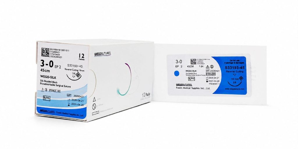
Material selection is the closure decision that gets least attention and produces the widest range of outcomes. A monofilament works differently from a braided suture in a contaminated environment. An absorbable choice degrades on a timeline that may or may not match the tissue healing curve. Choosing a surgical suture without matching the material to the site is the most common error in oral surgery, and it usually appears not as a dramatic failure but as slower healing that the clinician attributes to patient factors.
Silk is comfortable to handle but wicks bacteria into the wound, which matters in the oral cavity where the fluid environment is never sterile. PTFE monofilaments resist bacterial adherence and produce cleaner tissue response, at the cost of slightly less forgiving handling characteristics. Polyglycolic acid absorbables provide reliable timeline predictability but degrade faster in the alkaline conditions of some oral wounds. The right choice depends on procedure type, patient hygiene, and expected removal date, and it deserves a moment of deliberate thought rather than a reflexive habit.
Size Selection That Actually Matches the Tissue
Oversized sutures traumatize tissue, extend healing time, and produce visible marks. Undersized sutures fail under tension and reopen wounds during the first day of healing. A four-zero suture suits most oral surgical closures; five-zero or six-zero fits finer areas like anterior papilla reconstruction. Using the same size across every case, regardless of tissue thickness, guarantees suboptimal outcomes in at least one direction. Stocking three sizes and choosing deliberately per case is not a burden; it is a five-second decision that compounds over hundreds of procedures.
Technique Errors That Look Correct
A knot that holds during the procedure can still fail the healing process. Tension is the single most influential variable in whether a closure supports or obstructs healing. Sutures tied under excessive tension strangulate the tissue edges, cutting off the microvascular supply that drives repair. The closure looks tight and clean at the end of surgery, then produces necrotic edges that dehisce within seventy-two hours. The correct tension approximates tissue without blanching it, which is a tactile judgment that requires attention rather than automation.
Needle Handling and Tissue Trauma
The needle pass creates a channel that either heals cleanly or becomes a slow-inflammation site. Grasping the needle at the wrong point along its curve, using needle holders with worn jaws, or reversing direction mid-pass all traumatize tissue in ways that add up. The needle should enter perpendicular to the tissue and follow its curve without deflection. Small habits like resetting the grip on the needle holder between passes reduce wear on the needle and produce cleaner entry points.
Knot Placement That Compromises Healing
The knot itself should sit to the side of the incision line, not directly over it. A knot placed over the wound produces localized pressure, delays epithelialization, and increases patient discomfort during the first week. Sliding the knot to the buccal or lingual side of the closure takes one additional deliberate motion during tying and produces meaningfully better healing. This is the kind of small technique refinement that experienced surgeons often demonstrate without explaining, which leaves newer clinicians missing the point entirely.
Removal Timing and Post-Operative Errors
Sutures left in place too long become foreign bodies that sustain inflammation past the point where they served a purpose. In most oral surgical contexts, non-absorbable sutures should come out at seven to ten days. Absorbable choices that persist beyond three weeks in the oral environment often indicate that the wrong material was selected for the site. Scheduling the removal appointment before the patient leaves the surgical visit prevents the drift that keeps sutures in place until the patient notices them.
Patient instruction is where good closure work is often undone. Rinsing too vigorously, brushing over the surgical site, or eating hard foods against the closure line produces mechanical failure that has nothing to do with the technique. Written instructions with specific timelines for each activity, given alongside verbal explanation, dramatically reduce the frequency of these post-operative issues. Assistants who reinforce the same message at the discharge stage build a redundancy that catches patients who tuned out during the doctor’s explanation.
Building a Closure Standard That Holds
A practice that reviews closure outcomes in monthly case discussions learns faster than one where each surgeon relies on personal memory. Photographing the closure at the end of each surgical case, then comparing it to the seven-day follow-up, produces a visual record that reveals patterns invisible to memory alone. This is not a demand for elaborate documentation; a smartphone photo takes fifteen seconds and provides months of useful reference material.
Standardizing the closure tray across surgeons in a group practice also reduces error frequency. When each surgeon uses different suture materials and needle types, the assistant has to remember individual preferences and inevitably makes substitutions during busy days. A common baseline tray, with individual preferences added as documented deviations, produces cleaner workflows and fewer improvised choices during procedures. Suppliers like Plovio provide procedure-specific suture kits that support this standardization approach for practices building consistent protocols.
Closing the Feedback Loop on Wound Closure
The errors that delay healing are rarely dramatic, and that is why they persist. A suture material chosen out of habit, a knot tied slightly too tight, a placement that sits over the incision line instead of beside it, or a removal appointment that drifts by a week each contribute small amounts to outcomes that a busy clinician attributes to other factors. Addressing them requires deliberate attention rather than heroic effort, and the payoff shows up in reduced complications and steadier healing across the practice.
Start with material selection, then examine tension, needle handling, and knot placement in sequence. Track outcomes with simple photo documentation and review them monthly. Standardize the closure tray where practical and involve the surgical assistant in maintaining the protocol. Each of these steps takes minutes to implement, and together they eliminate the quiet errors that undermine surgical work at exactly the moment the case seems finished.